Your Brightening Serum Is Fine. It Is Being Applied to the Wrong Disease.
'Dark spots' bundles at least five distinct processes that sit at different depths, respond to different things, and have wildly different prognoses — one clears on its own, one relapses forever, and one will not move for any serum ever made.
Read the preview
From the DermApproved blog, in Hyperpigmentation: the red mark left behind by a spot is not pigment. It's blood vessels — so every brightening ingredient ever made is aimed at a pathway that mark isn't using. People spend a year on it and decide their skin doesn't respond. There's a three-second test that tells you which you have. For the full breakdown, visit dermapproved.com/blog.
One shelf, five diseases
The brightening aisle is organised around a symptom rather than a diagnosis, which is unusual in medicine and would be considered odd anywhere else. Imagine a pharmacy section labelled 'pain' containing one product.
At minimum, five things are being sold to at once. Post-inflammatory hyperpigmentation is brown pigment left where inflammation happened — a spot, a scratch, an eczema patch — and it fades on its own given time. Post-inflammatory erythema is the red or purple mark left after the same event, and it is not pigment at all. Melasma is symmetrical patchwork pigment on the cheeks, forehead and upper lip, hormonally and light-driven, with a strong tendency to relapse. Solar lentigines are the discrete, sharply defined brown patches that accumulate on chronically exposed skin with age. Ephelides — freckles — are genetically determined, appear early, and darken and fade seasonally with sun exposure.
They look similar enough on a phone camera to be confused, and they behave so differently that treating one as another wastes months. The most consequential distinction is not what caused them but how deep the pigment sits, because that single variable predicts whether anything you can apply will work at all.
Depth decides what is possible
Melanin deposited in the epidermis sits within reach of topical agents, is turned over as keratinocytes migrate upward, and will fade — slowly, but genuinely. Melanin that has dropped into the dermis, usually through damage to the dermo-epidermal junction during inflammation, is a different situation entirely: it is taken up by dermal macrophages, has no turnover conveyor to ride, and can persist for years. A review of post-inflammatory hyperpigmentation in skin of colour puts it plainly — topical therapy is typically effective for epidermal PIH, whereas recalcitrant disease may need chemical peeling or laser.
Clinically, epidermal pigment tends to be a well-demarcated brown; dermal pigment reads greyer, bluer or more muted, with less crisp edges. Wood's lamp examination is the traditional way of separating them, because epidermal pigment becomes more sharply contrasted under it while dermal pigment does not. This is worth knowing because it converts an open-ended question — how long until this goes — into a bounded one.
The implication people find hardest to accept is that a serum has a ceiling defined by anatomy rather than by formulation quality. A better tyrosinase inhibitor does not acquire the ability to reach the dermis. When a mark has not moved after six months of a good routine, the useful hypothesis is not that the routine was insufficiently expensive. It is that the pigment is somewhere the routine cannot go, and the hyperpigmentation pathway sets out what the procedural options actually are at that point.
The mark that is not pigment
Here is the mistake that costs the most time, and it happens overwhelmingly to people treating acne marks.
After a spot resolves it can leave brown pigment or it can leave a flat red-to-purple mark. The second is post-inflammatory erythema: dilated or damaged capillaries in the papillary dermis, a vascular finding with no melanin involvement. Every tyrosinase-inhibiting active ever formulated — hydroquinone, arbutin, kojic acid, vitamin C — is aimed at a pathway that is not operating in that mark. You can apply them faithfully for a year with no effect, and the reasonable conclusion drawn is that the skin is unresponsive, when the drug was simply aimed at the wrong tissue.
The practical test is embarrassingly simple: press on the mark, or stretch the skin. Erythema blanches because you have pushed blood out of the vessels; pigment does not. Thirty seconds, no equipment, and it redirects the entire plan. Treatment for the vascular version leans on time, on not re-inflaming the area, and where intervention is wanted, on vascular targets — a split-face study comparing intradermal tranexamic acid with pulsed dye laser for post-acne erythema found comparable efficacy between the two, which tells you what class of intervention the problem actually belongs to.
For genuine post-inflammatory pigment, the actives do work, and the ones with the best-established place are the unglamorous ones. Azelaic acid in something like The Ordinary Azelaic Acid Suspension or Naturium Azelaic Acid Emulsion is well tolerated on reactive skin. Niacinamide paired with tranexamic acid, as in Good Molecules Discoloration Correcting Serum, is the low-irritation option. And a retinoid such as Differin Adapalene Gel earns its place by treating the acne that keeps generating new marks, which matters more than fading the old ones.
Melasma plays by different rules
Melasma is the one that breaks routines built for everything else, because it is not primarily a damage-response problem. It is chronic, it has a hormonal dimension, it frequently has a dermal component, and it relapses. Framing it as something to cure sets up a failure; framing it as something to control makes the management legible.
The single most useful modern finding concerns what triggers it. Irradiation experiments on volunteers with skin types IV to VI compared long-wavelength UVA against visible light and found that visible light produced pigmentation that was both darker and more sustained — and that in skin type II, no pigmentation was observed at all. Visible light passes through window glass, is emitted by the sun on overcast days, and is entirely unaddressed by a conventional broad-spectrum sunscreen. For someone with melasma and deeper skin, that is not a footnote; it is most of the problem.
And it has a testable product consequence, which someone tested. Sixty-eight melasma patients all received hydroquinone 4%, randomised to either an iron-oxide-containing UV-plus-visible-light sunscreen or a UV-only sunscreen at SPF 50 or above. At eight weeks the iron oxide group showed 15% greater improvement in MASI, 28% greater improvement on colorimetry, and less melanin on histology. Same drug, different sunscreen, materially different outcome. A separate randomised trial found sunscreen covering short-wavelength visible light reduced melasma relapses.
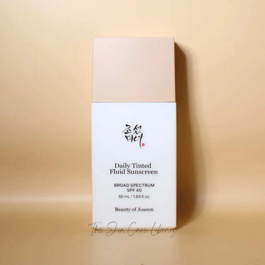
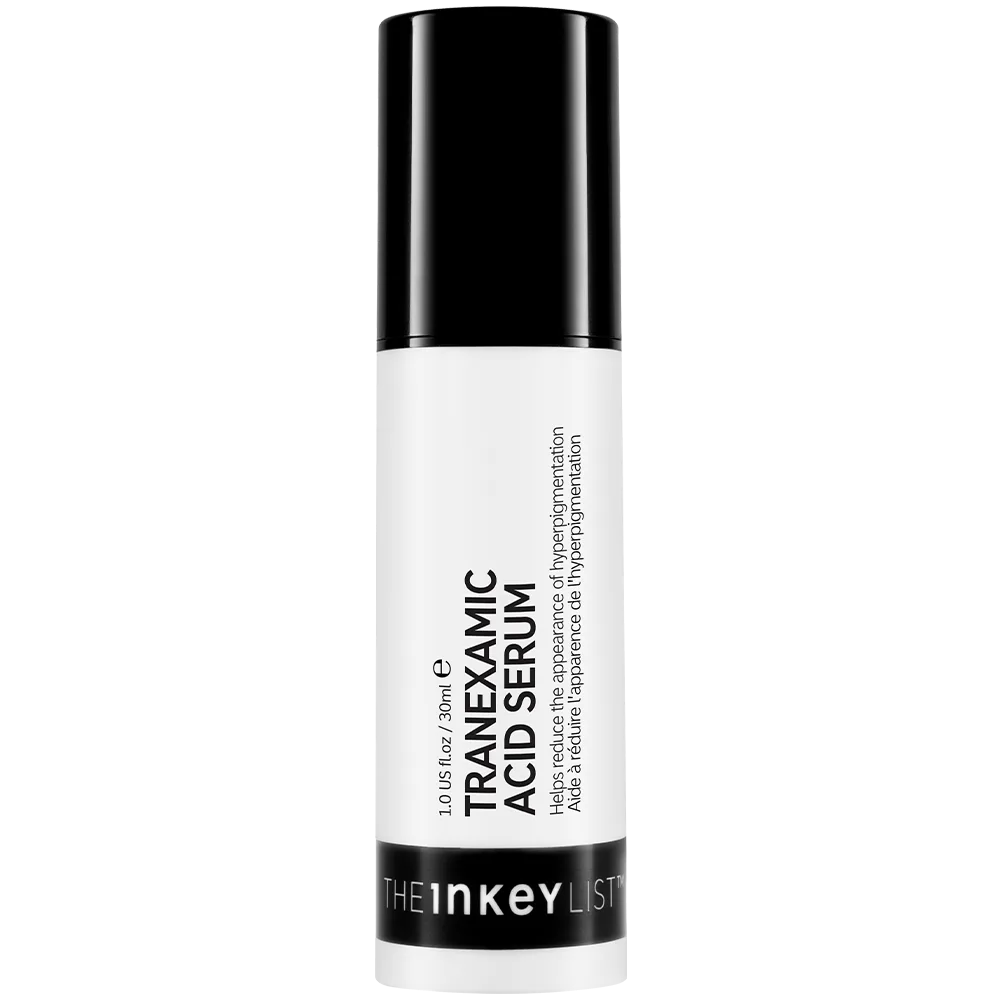
So the tinted sunscreen recommendation for melasma is not a cosmetic preference — the tint is the active pigment-blocking component. La Roche-Posay Anthelios Mineral Tinted SPF 50, ISDIN Eryfotona Ageless Tinted SPF 50 and Beauty of Joseon Daily Tinted Fluid SPF 40 are all doing something an untinted sunscreen structurally cannot. On the treatment side, tranexamic acid has become a mainstay — The INKEY List Tranexamic Acid Night Treatment topically, with the oral form a prescription conversation — and the reference regimen remains the triple combination of hydroquinone, a retinoid and a low-potency corticosteroid, which is a clinic decision rather than a shelf one. Because melasma waxes and wanes, tracking it formally is more useful than in any other pigment problem: the MASI Calculator gives you the score the trials use, and the Progress Photo Tracker keeps the lighting consistent enough that the comparison means something.
The spots that will not move
Solar lentigines are where most brightening routines quietly fail, and where the honest advice is to stop buying serums.
These are the sharply demarcated brown patches on the backs of hands, forehead, upper chest and cheeks that accumulate with cumulative exposure. Histologically they involve increased melanocyte numbers and elongated rete ridges — a structural change in the epidermis rather than a transient surge in pigment production. A systematic review of 41 clinical trials covering 3,234 patients found the most effective topical option was a combination of mequinol and tretinoin, with lasers and intense pulsed light performing considerably better: intense pulsed light reported success in the region of three-quarters to ninety per cent of cases, picosecond lasers similarly high, while cryotherapy and trichloroacetic acid peels were less reliable and cryotherapy carried the worst side-effect profile.
The conclusion for anyone with a face full of these is unromantic. A retinoid such as The Ordinary Retinol 0.2% in Squalane and diligent sun protection will slow the accumulation of new ones and modestly improve the surrounding skin. They will not clear an established lentigo, and neither will a vitamin C serum, however good. The intervention that removes them is a device, and knowing that early is worth more than a drawer of half-used bottles. Freckles sit in a similar bracket for different reasons: genetically determined, sun-responsive, and best managed by not chasing them.
What unites all five, and the only universal instruction in this piece, is photoprotection. Every one of these conditions is driven or aggravated by light, every treatment for them is undermined by continued exposure, and every procedure that removes pigment can itself trigger post-inflammatory pigment if the skin is subsequently unprotected. The Sun Safety Guide covers the boring specifics — quantity, reapplication, what shade actually does — and the pigmentation routine assembles the rest into an order you can follow. Identify the disease first. The serum is not the problem.
Products mentioned
Independently reviewed and scored — the real products behind this piece.




- Therapy guide Hyperpigmentation What the procedural options are once topical therapy has hit its anatomical ceiling.
- Routine Skincare Routine for Hyperpigmentation The actives assembled into an order, once you know which disease you are treating.
- Ranked list Best Sunscreens for Hyperpigmentation Screened on the criterion that actually matters here, including visible-light coverage.
- Ranked list Best Azelaic Acid for Hyperpigmentation For the post-inflammatory pigment case specifically.
- Ranked list Best Tranexamic Acid for Hyperpigmentation For the melasma lane, where tranexamic acid has become standard.
- Ingredient Azelaic Acid Well tolerated, effective on epidermal pigment, and usable where retinoids are not.
- Ingredient Niacinamide (Vitamin B3) Interferes with melanosome transfer rather than melanin synthesis — a different lever.
- Ingredient Vitamin C (L-Ascorbic Acid) A tyrosinase-directed active, and therefore useless against the red post-acne mark.
References
- Postinflammatory hyperpigmentation: a review of the epidemiology, clinical features, and treatment options in skin of color — The Journal of Clinical and Aesthetic Dermatology, 2010
- Impact of long-wavelength UVA and visible light on melanocompetent skin — Journal of Investigative Dermatology, 2010
- Near-visible light and UV photoprotection in the treatment of melasma: a double-blind randomized trial — Photodermatology, Photoimmunology & Photomedicine, 2014
- Prevention of melasma relapses with sunscreen combining protection against UV and short wavelengths of visible light: a prospective randomized comparative trial — Journal of the American Academy of Dermatology, 2015
- Treatment of Solar Lentigines: A Systematic Review of Clinical Trials — Journal of Cosmetic Dermatology, 2025
- The Comparable Efficacy Between Tranexamic Acid Intradermal Injection and Pulsed Dye Laser for Treatment of Post-Acne Erythema: A Split-Face Therapy — Clinical, Cosmetic and Investigational Dermatology, 2025
- Efficacy and safety of a new triple-combination agent for the treatment of facial melasma — Cutis, 2003
Every cited link is verified to resolve to the paper named. Where a source could not be independently confirmed, it is listed without a link rather than pointing you somewhere wrong.
Frequently asked
How do I tell a brown acne mark from a red one, and does it matter?
Press on it or stretch the skin. A red or purple mark blanches because it is dilated vasculature; pigment does not change. It matters enormously — every tyrosinase-inhibiting active is aimed at melanin production, so applying them to post-inflammatory erythema means treating a pathway that is not involved.
Why is a tinted sunscreen specifically recommended for melasma?
Because the tint is functional. Visible light induces pigmentation in deeper skin types — in controlled irradiation it produced darker and longer-lasting pigment than long-wave UVA — and conventional sunscreens do not filter it. In a randomised trial, adding an iron-oxide sunscreen to hydroquinone produced 15% greater MASI improvement over eight weeks than the same drug with a UV-only sunscreen.
My dark spots haven't budged in six months. Should I switch to a stronger serum?
Probably not. Consider instead that the pigment may sit in the dermis, where no topical reaches, or that the lesions are solar lentigines, which are a structural epidermal change rather than a transient pigment surge. Both scenarios have the same answer, and it is a procedure rather than a bottle.
Do solar lentigines ever respond to topical treatment?
Modestly and slowly. In a systematic review of 41 trials, the most effective topical was a mequinol-plus-tretinoin combination, while intense pulsed light and picosecond lasers reported far higher success rates. Retinoids and sun protection are worth using to slow new ones appearing; established spots generally need a device.
Will post-inflammatory hyperpigmentation go away on its own?
Epidermal pigment usually does, over months, provided the underlying inflammation is controlled and the area is protected from light. Dermal pigment can persist for years. Treating the original condition — the acne, the eczema, the friction — matters more than any fading agent, because new marks form faster than old ones clear.